
The 18 parts of the leg (characteristics and functions)
The human body is much more than the mere sum of the 30 million million cells that make it up. It is an almost perfect machine and an absolute feat of biological evolution in which the different organs and tissues of the body are coordinated in an amazing way to allow us to fulfill our physiological and mechanical functions.
And although when we talk about characteristics that have made the human being something unique in nature, we generally think of the brain, this is something unfair to the other great characteristic that differentiated us from other animals: bipedalism. Walking on only two limbs is a trait that, despite being so integrated into our mentality today, in its day marked an enormous evolution within the animal kingdom.
And in this context, bipedalism is possible, among many other things, thanks to the evolution of the legs, the lower extremities of our body, which have adapted to allow this standing and make it possible to walk, jump and run. Structures that, although not vital for life, are practically essential for living.
Thus, in today's article and, as always, from the hand of the most prestigious scientific publications, we are going to review the morphology and structure of the legs, analyzing the bones and muscles that make up the lower extremities of our organism. Let us begin.
What is the morphology of the legs?
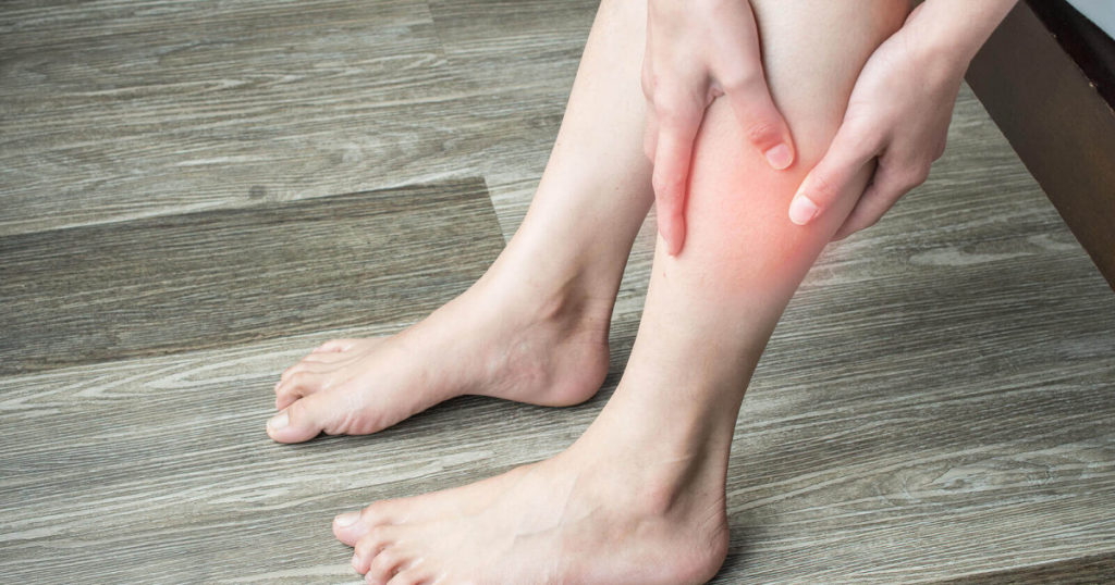
The legs are, at the technical level of human anatomy, the region between the knee and the ankle, thus being known as the third segment of the lower limb. It is the anatomical area that articulates with the thigh through the knee and with the foot through the ankle. Even so, at the level of popular culture, we understand the leg as the totality of the lower extremities.
For this reason and to offer the maximum possible information, although we do not fit so much in the exact definition, we are going to explore the parts that make up the legs, understanding them as the entire lower limb. Let's see, then, by what morphological structures a human leg is constituted and what are its anatomical characteristics and physiological functions.
1. Leg bones
The bones are the mainstay of the locomotor system and, of course, the bony component of the legs is essential in its morphology. The legs are made up of three main bones (we could also speak of the patella, which is a flattened bone exclusive to the knee): femur, fibula, and tibia. Let's see the characteristics, location and functions of each of them.
1.1. Femur
The femur is the longest and strongest bone in the human body. It extends from the thigh and is relatively spherical in the most distal part of the thigh to fit the knee, the largest joint in the human body. Therefore, the femur is the leg bone above the knee.
1.2. Fibula
The fibula is one of the bones that, together with the tibia, constitutes the bony component of the region of the lower trunk below the knee. Of these two bones below the knee joint, the fibula is the least voluminous and is located on the outer side (the part furthest from the other leg), allowing the lower trunk to articulate by connecting with the knee.
1.3. Tibia
The tibia is the bone that it is close to the fibula, being, of the two, the largest and most voluminous. It is located on the inside of the leg (the part closest to the other leg) and in an anterior region, that is, in front of the fibula. Like this, its function is to, through the connection with the knee, make possible the articulation of the lower trunk.
2. Leg muscles
Muscles are the organs of the locomotor system that, being made up of muscle tissue and connected to the nervous system, have the ability to contract and relax in order to transmit movement to the bones and make locomotion possible. And, of course, the muscular component of the legs is very important. These are the main muscles of the lower extremities.
2.1. Quadriceps
The quadriceps is a muscle made up of four parts (vastus medialis, vastus lateralis, vastus medialis and rectus anterior) that is located in the anterior part of the thigh, originating in the hip and extending to the tibia, and that is designed to generate movements in the knee, support the weight of the body and allow us to walk, sit, run and jump. It is considered the strongest muscle in the human body.
2.2. Hamstrings
The hamstring muscles are a group of three muscles (biceps femoris, semitendinosus, and semimembranosus) that run along the back of the thigh from the hip to the region just below the knee. They are essential to allow knee flexion and leg extension.
23. Abductors
The abductors are six muscles (gluteus maximus, sartorius muscle, tensor fascia lata, pyramidal muscle, gluteus minus, and gluteus medius) located on the outside of the thigh and gluteus with the very important function of allowing the separation of the legs. That is, they allow us to raise the leg laterally and move it away from its axis, that is, from the midline of the body.
2.4. Adductors
The adductors are five muscles (pectineus muscle, adductor magnus, short adductor muscle, long adductor muscle, and gracilis muscle) located on the inner thigh and that are antagonistic to the abductors, since they have the function of closing the legs. They bring one leg closer to the other.
2.5. Anterior tibial
The tibialis anterior muscle is a thick muscle located in the anterior part of the leg, originating in the upper two-thirds of the external aspect of the tibia and ending in the first metatarsals of the foot. It has the important function of stabilizing the ankle.
2.6. Long extenders
The long extensor muscles are two muscles that are located in the anterior part of the leg, above the tibialis anterior muscle and that have the function of provoke the extension movement of the phalanges of the feet. There is one muscle that takes care of all the fingers except the big toe and one that only takes care of this toe.
2.7. Anterior peroneum
The peroneus anterior is a small muscular belly that, located in the anterior part and the external aspect of the leg, arises in the lower third of the fibula and has the function of causing dorsal flexion of the foot and eversion, which is the elevation of the outer edge so that the plant of the same one looks out.
2.8. Lateral fibulae
The lateral peroneal muscles are two muscles that are found on the outer and lateral surface of the leg. We have the peroneus longus and peroneus brevis. The length originates from the external tuberosity of the fibula and has the function of extending the foot over the leg, bringing it out and executing a rotary movement. The short has the function of causing abduction (separating from the axis) and external rotation of the foot.
2.9. Popliteal
The popliteal is a muscle located at the back of the knee and that, being triangular, flattened and short, is the flexor of the leg on the thigh and medial rotator of the knee when the knee is in flexion. By internally rotating this joint, you unlock it when walking or running.
2.10. Long flexor of the toes
The long flexor of the toes is a muscle originating in the middle part of the posterior aspect of the tibia and that allows the flexion of the toes, that is, they make possible the movement by which we bring the foot closer to the leg parallel to the sagittal plane.
2.11. Posterior tibial
The tibialis posterior is an elongated muscle located at the back of the leg, originating from the proximal part of the tibia. It has three main functions: foot adduction (the sole of the foot faces inward), plantar flexion, and stabilization of the plantar arch (the dome-shaped structure on the soles of the feet).
2.12. Gastrocnemius
The gastrocnemius is a muscle separated into two halves and, located at the back of the leg, is the most superficial muscle of the calf. Its function is to slightly contribute to the flexion of the knee but, above all, to make plantar flexion of the foot possible. Popularly known as the twin, it is essential to give propulsion at the beginning of the march.
2.13. Soleus
The soleus is a thick and broad muscle located on the back of the leg, below and behind the calf. In fact, they are so closely related that many experts believe that only one muscle should be discussed: the triceps surae. In any case, this soleus has the importance of allowing, in addition to the plantar flexion and extension of the foot, the elevation of the heel.
2.14. Plant thin
The plantar slender is a muscle located at the back of the leg, although it has a variable size and extension and some people may not even present it. It lies deeper than the gastrocnemius and its motor action, despite contributing to flexion of the foot and knee, is considered quite weak.
2.15. Long flexor of the big toe
The flexor hallucis longus is a muscle located at the back of the calf, deeper than the gastrocnemius. It inserts into the lower third of the fibula and, by contracting, makes it possible to flex the big toe.
Leave a Reply