The 3 types of cartilage (and their characteristics)
Human beings are much more than the sum of the 30 million million cells that make up our body. We are a feat of biological evolution in which these cells differentiate and organize themselves into different cell types with specific morphological and physiological characteristics to fulfill specific purposes in the body.
And it is in this context that tissues come into play, a concept that designates the set of cells with a similar pattern of genetic expression organized among them forming an anatomically complex structure that, in turn, gives rise to the different organs of the body. And it is from the combination of 14 cellular tissues that our morphological and functional diversity emerges.
Some tissues are the first that come to mind when we think about them, such as epithelial tissue, blood, nervous tissue, muscle tissue, bone tissue or adipose tissue, but there are others that, despite being Just as important, they go unnoticed. And one of them is undoubtedly cartilaginous tissue.
Cartilage tissue is that which makes up, as its name suggests, the cartilage of the body. Connective tissue structures that prevent friction between the bone pieces of a joint and that, in turn, shape different regions of the body such as the ears, nose or trachea. And in today's article we will analyze the characteristics and classification of these cartilages.
What is a cartilage?
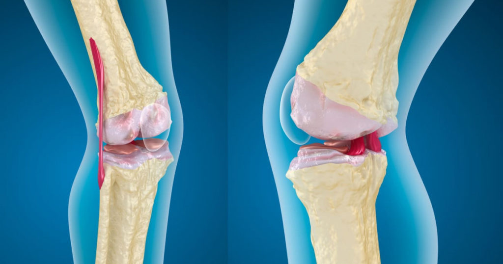
A cartilage is a structure of cartilaginous tissue that prevents friction between the bony parts of the body's joints and, in turn, shapes different regions of the body such as the ears, nose or trachea. It is a type of connective tissue rich in chondrogenic cells, collagen and elastic fibers, thus being very resistant structures essential in the joints.
As we say, this cartilage tissue is a type of connective tissue, also known as connective tissue, which means that its cells are designed to hold other tissues and organs together, mechanically and physiologically connecting them. There are many different connective tissues (such as blood), since all those that "fill in" the spaces between tissues, that keep the organs in their position and make sure that the body has its proper shape, are connective tissues.
But one specific type is this cartilaginous tissue. We are facing an elastic tissue formed mainly by an extracellular matrix and by specific cells of the same. On the one hand, this extracellular cartilaginous matrix is made up of type II collagen (collagen is a protein that holds different structures of the body together, and in this specific case forms fine fibrils), type IX collagen (one of the type II collagen fibrils). II to each other), type X collagen (surrounds cells in a hypertrophic state), type XI collagen (its function remains unclear) and hyalurane, which, together with the proteoglycan aggregates that bind to it, is responsible for the typical cartilaginous consistency.
This extracellular matrix is what gives cartilage its strength, stability and consistency, but we cannot forget its cellular component.. In "gaps" in this cartilaginous matrix (technically known as chondroplasts) are located the chondrocytes, which are the cells that, being dispersed, make up the cellular component of the cartilaginous tissue and synthesize the matrix.
However, it must be taken into account that mature cartilage lacks both blood and nerve supply, which is why it is neither colored nor sensitive, respectively. Therefore, by not being able to receive nutrients through the blood, these chondrocytes "feed" through a diffusion process through the matrix, developing an anaerobic metabolism in most cases.


Be that as it may, the important thing is that cartilages are organic structures of cartilaginous tissue (a class of connective tissue) that, although they are found in the embryos of vertebrates and cartilaginous fish, in the adult human body they develop their main function by coating the joints, positioned between the bone pieces to avoid friction between them during joint movement.
In fact, it is precisely on the cartilage that the synovial fluid typical of the synovial joints is deposited (those that, unlike the solid ones, allow movement) forming a layer 50 micrometers thick and penetrating inside so that, in When making a movement, this synovial fluid emerges from the cartilage and, like the oil we put on the hinges, keeps the joint lubricated.
Even so, we must bear in mind that cartilage cannot regenerate. Hence, its wear is progressive and chronic and that, at a point where this degeneration is enough for the bones of a joint to rub against each other, disorders such as osteoarthritis may appear, which causes pain when moving and joint deformity.
But the role of cartilage is not limited exclusively to the joints, where it prevents friction wear and cushions blows. We also have cartilage in the trachea and bronchi, reinforcing these structures, in the outer ear (giving shape to what we traditionally understand as the ear), in the nasal septum and even in the joints between the ribs and the sternum. Cartilage, then, are essential in our body.
How is cartilage classified?
After this extensive but absolutely necessary introduction, the biological bases of cartilage tissue have surely become more than clear. In any case, the truth is that, depending on their morphological and physiological properties, cartilage can be classified into different groups. So let's see what types of cartilage exist.
1. Hyaline cartilage
Hyaline cartilage is the most abundant in our body, being the one that is present not only in the joints, but also in the nose, trachea, bronchi, larynx and the ventral ends of the ribs. It has a bluish-white appearance, has few fibers and has perichondrium, a dense, irregular, collagen-rich connective tissue sheath that covers this cartilage, except for the cartilage in the epiphyses, that is, the widened ends of the long bones. , and articular cartilages.
These hyaline cartilages stand out for their composition in type II collagen fibrils, for their chondrocytes (the cellular component of cartilage) organized in groups (known as isogenic groups, each one of them surrounded by a territorial matrix) and for their matrix basophilic, that is, it is easily stained with basic dyes. It is avascular, that is, it lacks blood supply, so the chondrocytes are nourished by diffusion through the synovial fluid.


2. Elastic cartilage
The elastic cartilage stands out, in addition to its special elasticity (something logical seeing its name), superior to that of the other two types, due to its yellowish coloration. It is present in the pinna, that is, it is the one that shapes the ear, in the epiglottis (the sheet-shaped organ that closes the upper opening of the larynx at the time of swallowing), in the Eustachian tube (tube that connects the middle ear with the pharynx), in the walls of the auditory canal and constituting the cuneiform cartilage of the larynx.
All elastic cartilages have the aforementioned perichondrium, that is, the collagen-rich connective tissue sheath that, being irregular and dense, is located covering the cartilage. It stands out for its composition in type II collagen fibrils and for its high amount of elastic fibers, which give it this flexibility that defines it. It is, like hyaline, always avascular, that is, it lacks blood supply.
It has a greater number of isogenic groups (organization of chondrocytes, the cellular component of cartilaginous tissue) and stands out mainly because its cartilaginous matrix presents a very dense interwoven of fine elastic fibers that, like hyaline cartilage, makes it basophilic, it is that is, it is stained with basic dyes.


3. Fibrous cartilage
Finally, fibrous cartilage, also known as fibrocartilage, is a type of cartilaginous tissue present in the insertion of some tendons (the bundles of collagen-rich connective fibers that connect muscles to bones), in the articular discs, the intervertebral discs (in the spine), the pubic symphysis, which is the connection between the two parts of the pubis, the menisci of the knees, the jaw, and essentially everywhere there is an intersection between ligaments (bundles that join bone to bone) and tendons.
All fibrous cartilages lack perichondrium, the fibrous membrane that surrounds all elastic cartilage and most hyalines. Its composition stands out for its type I collagen fibrils and because its matrix is acidophilic, that is, unlike the previous two, it is stained with acid, not basic, dyes.
In this fibrous cartilage, the chondrocytes organize themselves forming a kind of parallel rows between the collagen bundles. It is a transition between the regular dense connective tissue and the hyaline cartilage that we mentioned earlier. It is generally avascular, but there are exceptions of cartilage where there is blood supply. In addition, cartilage regeneration therapies do not work in this, as they are designed for hyaline cartilage. Hence, the treatment of a torn meniscus is particularly complex.


Leave a Reply